Human Lower Body Diagram : List Of Skeletal Muscles Of The Human Body Wikipedia / 674 x 599 photo description:. The basic parts of the human body are the head, neck, torso, arms and legs. Evenly distribute weights from your upper body into the lower extremities. Your lower back (lumbar spine) is the anatomic region between your lowest rib and the upper part of the buttock. The spine diagram shown below, consists of many bones or vertebrae,soft discs,the spinal cord, and spinal nerves. This makes the knowledge about human body is just as important as anything.
The majority of muscles in the leg are considered long muscles, in that they stretch great distances. The lower leg is a major anatomical part of the skeletal system. Diagrams of the human body. The femur is the largest bone in the body and the only bone of the thigh (femoral) region. The muscles of the abdomen protect vital organs underneath and provide structure for the spine.
Femur Wikipedia from upload.wikimedia.org The myology of the lower limb is also particularly well represented in this atlas of anatomy, with multiple anatomical charts and diagrams: Human internal organs vector vector isolated illustration of human internal organs in female body. The first diagram summarizes the different muscular compartments (fascial compartments) of the thigh and leg, and the different fascias (crural fascia, intermuscular septum, interosseous membrane, adductor canal, fascia lata) Glands in the human body produce, control and regulate the flow of hormones, breast milk, saliva, and other useful fluids. It contains the osteology, arthrology and myology of the spine and back. 674 x 599 photo description: The knee joins the upper leg and the lower leg. Formed by the left and right hip bones, the pelvic girdle connects the lower limb (leg) bones to the axial skeleton.
Do you ever wonder what the major organs of the body are and.
The lower leg is a major anatomical part of the skeletal system. This curve, called lordosis, helps to: The bones of the pelvis and lower back work together to support the body's weight, anchor the abdominal and hip muscles, and protect the delicate vital organs of the vertebral and abdominopelvic cavities. The major muscles of the abdomen include the rectus. For teachers, students, health professionals, or anyone interested in learning about the anatomy of the human body. Diagrams of the human body. The vertebrae, which stack like spools of thread, support the back and protect the spinal cord. Muscle charts of the human body for your reference value these charts show the major superficial and deep muscles of the human body. Stomach, liver, intestine, bladder, lung, testicle, uterus, spine, pancreas, kidney, heart. Together with the upper leg, it forms the lower extremity. The pain body map is part of an update of content on the website by the michael g. These muscles help the body bend at the waist. For more anatomy content please follow us and visit our website:
For more anatomy content please follow us and visit our website: It is particularly interesting for physiotherapists. This curve, called lordosis, helps to: Diagrams of the human body. We hope this picture human body artery diagram in detail can help you study and research.
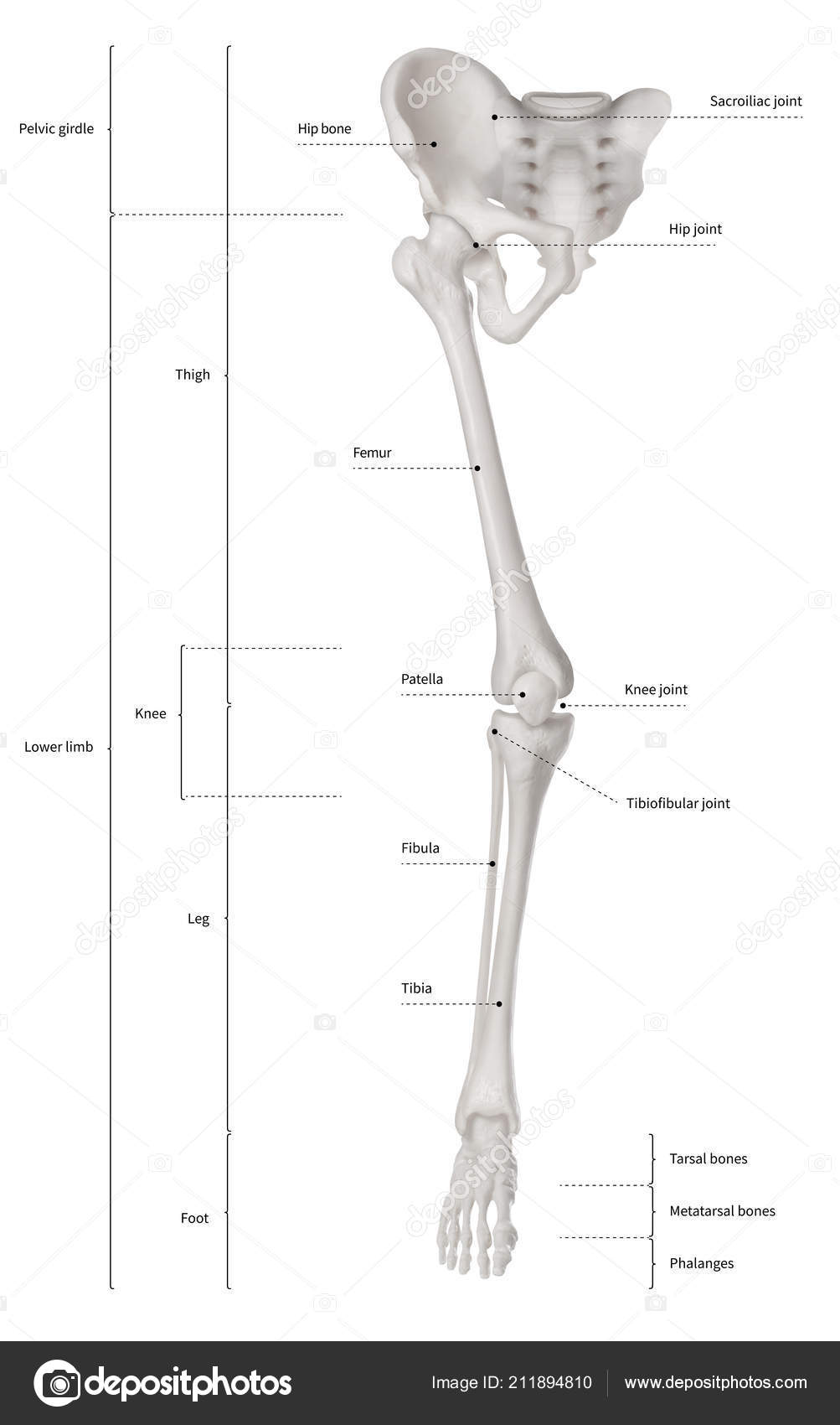
Infographic Diagram Human Skeleton Lower Limb Anatomy Bone System Leg Stock Photo By C K Intarapong Gmail Com 211894810 from st4.depositphotos.com The vertebral column of the lower back includes the five lumbar vertebrae, the sacrum, and the coccyx. This curve, called lordosis, helps to: 1 your spine in this region has a natural inward curve. The lower leg is a major anatomical part of the skeletal system. In turn, the spinal cord relays essential information between the brain and the body. Daniel nelson on june 5, 2018 8 comments ! For teachers, students, health professionals, or anyone interested in learning about the anatomy of the human body. Shop items you love at overstock, with free shipping on everything* and easy returns.
Anatomical diagrams of the spine and back.
674 x 599 photo description: Balance the weight of your head on top of your spine. This diagram depicts human body map of organs with parts and labels. Human organs & anatomy diagram picture category: The myology of the lower limb is also particularly well represented in this atlas of anatomy, with multiple anatomical charts and diagrams: Posted in diagrams scalenes muscles. It's also the largest joint in the body. The spine anatomy is a complex structure. In addition to bearing the weight of the upper body, the knee allows for walking, running, and jumping. Find free pictures, photos, diagrams, images and information related to the human body right here at science kids. Together with the upper leg, it forms the lower extremity. It lies between the knee and the ankle, while the upper leg lies between. This diagram depicts lower extremity diagram.human anatomy diagrams show internal organs, cells, systems, conditions, symptoms and sickness information and/or tips for healthy living.
The pain body map is part of an update of content on the website by the michael g. The muscles of the abdomen protect vital organs underneath and provide structure for the spine. Together with the upper leg, it forms the lower extremity. This article explains the significance of glands in metabolism, growth, and reproduction. The bones of the pelvis and lower back work together to support the body's weight, anchor the abdominal and hip muscles, and protect the delicate vital organs of the vertebral and abdominopelvic cavities.
Lower Body Anatomy Artwork Stock Image F005 9864 Science Photo Library from media.sciencephoto.com Diagram lower back wiring diagram advance together the brain and spinal cord make up the central nervous system. Daniel nelson on june 5, 2018 8 comments ! The myology of the lower limb is also particularly well represented in this atlas of anatomy, with multiple anatomical charts and diagrams: Do you ever wonder what the major organs of the body are and. The majority of muscles in the leg are considered long muscles, in that they stretch great distances. The diaphragm forms the upper surface of the abdomen. The sciatic nerve is the dominant nerve that innervates the lower back and the lower extremities. It is particularly interesting for physiotherapists.
The sciatic nerve is the dominant nerve that innervates the lower back and the lower extremities.
As these muscles contract and relax, they move skeletal bones to create movement of the body. The vertebral column of the lower back includes the five lumbar vertebrae, the sacrum, and the coccyx. At the level of the pelvic bones, the abdomen. The muscles of the lower back help stabilize, rotate, flex, and extend the spinal column, which is a bony tower of 24 vertebrae that gives the body structure and houses the spinal cord. In turn, the spinal cord relays essential information between the brain and the body. It contains the osteology, arthrology and myology of the spine and back. 1 your spine in this region has a natural inward curve. The muscles of the abdomen protect vital organs underneath and provide structure for the spine. This makes the knowledge about human body is just as important as anything. Anatomynote.com found human body artery diagram in detail from plenty of anatomical pictures on the internet. Learn anatomy as you browse our collection of colorful, large and clearly labeled human body diagrams. This diagram depicts lower extremity muscles diagram.human anatomy diagrams show internal organs, cells, systems, conditions, symptoms and sickness information and/or tips for healthy living. In all, there are believed to be 80 organs in your body, all serving different functions and uses.
Find free pictures, photos, diagrams, images and information related to the human body right here at science kids lower body diagram. Balance the weight of your head on top of your spine.


Posting Komentar
0 Komentar